animal cell under electron microscope labelled
You see that many features are in common. Cell And Organelles Dr Jastrow S Electron Microscopic Atlas.

Related Image Cell Diagram Plant Cell Diagram Plant Cell
Calculate the actual length of the organelle as shown by the line AB in the diagram.
. There are one or more cells that form organism. A click on eg Blood opens an overview page on the different blood cells. To make observations and draw scale.
A cell is the smallest functional and structural entity of life that it is easier observing animal cell under light microscope. Ii Which parts are concerned with the following functions. This is a close up of a fly.
There are two categories of cells Eukaryotic and Prokaryotic. During mitosis the two sets of chromosomes are precisely separated and each daughter cell receives one complete set. Sep 15 2014 - Learn the structure of animal cell and plant cell under light microscope.
A B C D 4 iiStructures C and E are examples of the same organelle. See how a generalized structure of an animal cell. Sel Tumbuhan Dan Sel Hewan Cell Organelles Animal Cell Cell Diagram.
Ib Biology 1 2 Slides Ultrastructure Of Cells. This is a colored scanning electron micrograph of human red and white blood cells. The electron micrograph displayed below illustrates many of the plant cell characteristics discussed The cell wall large central vacuole and chloroplasts are clearly visible Also visible is the clearly defined nucleus containing chromatin Nucleus Chromatin The vacuole in this mature plant cell from a leaf is large and occupies about 80 of.
Generalized Structure of Animal Cell Plant Cell Under Microscope Generalized Structure of a Plant Cell Diagram As you can see in the above labeled plant cell diagram under light microscope there are 13 parts namely Cell membrane Cytoplasm Ribosomes Nucleus Smooth Endoplasmic Reticulum Lysosome Chloroplast Cell Wall Vacuole Golgi bodies. Cell is a tiny structure and functional unit of a living organism containing various parts known as organelles. Compare an animal cell to a plant cell.
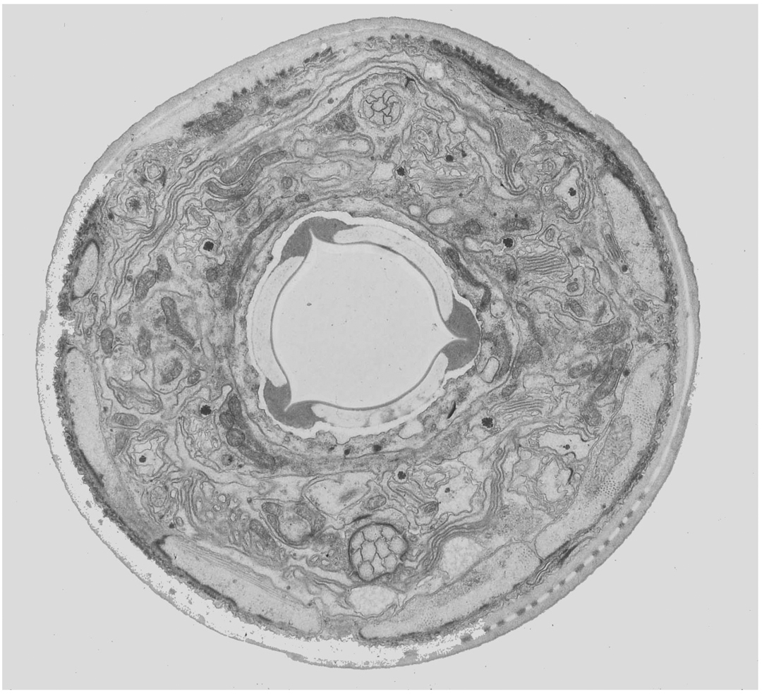
The figure below is a diagram of an animal cell as seen using a transmission electron microscope. Electron Micrograph Animal Cell Under Electron Microscope. So it is important to note that what we are drawing is definitely not life.
The plant cell as more rigid and stiff walls. In the given figure of an animal cell as observed under an electron microscope. The Figure Below Is A Fine Structure Of A Generalized Animal Cell.
This is a cancerous human skin cell. Tuesday April 20th 2021. Biologists gener Continue Reading Vern Shellman.
Animal Cell Diagram Under Light Microscope. Typical Animal Cell Pinocytotic vesicle Lysosome Golgi vesicles Golgi vesicles rough ER endoplasmic reticulum Smooth ER no ribosomes Cell plasma membrane Mitochondrion Golgi apparatus Nucleolus Nucleus Centrioles 2 Each composed of 9. Eukaryotic is most complex cells consisting a true nucleus enclosed by a membrane.
We all keep in mind that the human physique is amazingly elaborate and one way I discovered to comprehend it is by way of the style of human anatomy diagrams. This appears at the light microscope level as a duplication of chromosomes. Electron Microscope Eukaryotic Animal Cell Micropedia Labelled Plant Cell Microscope Image Micropedia Learn the structure of animal cell and plant cell under light microscope.
Cell Membranes And Compartments Cellbiology. Draw and label an animal cell as seen under an electron microscope. 25 Animal Cells Diagram With Labels Markcritz Template Design Cell Diagram Cell Diagram Project Plant And Animal Cells.
Before cell division the entire genome is copied. Bookfanatic89 Diagram Of Plant Cell Under Electron Microscope. A typical animal cell as seen in an electron microscope Medical Images For PowerPoint 1.
Here is an electron micrograph of an animal cell with the labels superimposed. Frontiers Soil Microstructures Examined Through Transmission. The animal cell is more fluid or elastic or malleable in structure.
Draw A Well Labelled Diagram Of A Plant Cell As Seen Under Electron Microscope. Fillable Online Diagram For Labelling Parts Of Plant. Animal cells have a basic structure.
I Name the parts labelled as 1 to 10. IName the structures of the cell labelled A B Cand D. Parts Of Microscope With Their Functions And Working Principle Electron Microscope Technology Life Microscope Parts.
Elodea Plant Cell Microscope Labeled. These are both specific types of cells and from specific species. Below the basic structure is shown in the same animal cell on the left viewed with the light microscope and on the right with the transmission electron.
Endoplasmic Reticulum Rough And Smooth British Society For. It was not until good light microscopes became available in the early part of the nineteenth century that all plant and animal tissues were discovered to be aggregates of individual cells. Table D leads to images of electron microscopes or protocols for tissue preparation.
Nucleus Plant Cell Microscope Labeled. Now the first thing to point out when looking at images under an electron microscope is the scale. Suggest why E looks so different to C.
Animal and plant cells undergo a precise type of division called mitosis. The structures within the cell are referred to as organelles. For example something that you draw as 3cm long may in fact be 10 000 times smaller in real life.
A typical animal cell is 1020 μm in diameter which is about one-fifth the size of the smallest particle visible to the naked eye. Labelled Plant Cell Under Microscope. Table E leads to the overview pages with the images of this atlas which are used in the histology course of the University of Mainz Germany.
A Release of energy b Protein synthesis c Transmission of hereditary characters from parents to. Eukaryotes prokaryotes and measuring cells vbiology labeled blood cells under microscope blood smear i electron microscopic structure of a typical bacterial cell liquid scanning transmission electron microscopy imaging microscopy cells scanning electron microscopy of cells and tissues under animal cell structure incl histology organelles. Pin By Simparinka Samuel On Bikotwa Junior Cell Diagram Plant Cell Diagram Plant Cell.
Animal Cell Diagram Under Electron Microscope. Science Aid Cell Structure Animal Cell Structure Cell Structure Cell Diagram. This shows a generalized animal cell under a light microscope.

560 X 364 Pixel Electron Microscope Image Animal Cell And Organelles Labeled Animal Cell Plasma Membrane Organelles

Histolab4a Htm Teaching Biology Cell Biology Plasma Membrane

What Causes Cancer Part Ii Mitochondria And Cancer Diagnosis Diet Cell Diagram Animal Cell Cells Worksheet

Structure Of Animal Cell And Plant Cell Under Microscope Diagrams Cell Diagram Plant Cell Diagram Animal Cell

Animal Cells Animal Cell Cell Biology Cell Model

Pin By Mustafa Tasyurekli On Sebzeler Animal Cell Structure Cell Organelles Electron Microscope

Pin By Nia On Biologie In 2021 Plant Cell Electron Microscope Cell

An Electron Micrograph Of A Mouse Liver Cell Dna Learning Center Electrons Cell Learning Centers

Cell Nucleus Function Structure And Under A Microscope Rs Science In 2021 Cells Lesson Cells Activity Things Under A Microscope

Microscopic Description Case 156 Animal Cell Organelles Cell Organelles Organelles

Year 11 Bio Key Points Cell Organelles And Their Function Cell Organelles Animal Cell Organelles

Plant Cell Diagram Animal Cell Diagram Plant And Animal Cells Science Cells Animal Cell

Pin By Katie Riley Peterson On Teaching Biology Animal Cell Animal Cell Structure Cell Diagram
Cell Biology Biology Animal Cell

This Schematic Diagram Shows A Generic Animal Cell And The Organelles Including The Nucleus En Animal Cells Worksheet Human Cell Diagram Human Cell Structure

Cell Theory Plant Cell Diagram Cell Diagram

Animal Cell Structure And Organelles With Their Functions Animal Cell Organelles Cell Diagram

What Is Going On Inside That Cell Cell Diagram Human Cell Diagram Human Cell Structure

Animal Cell Organelles Sauna Design